Health & Medicine
Living with a rare genetic disorder

Using a miniscope to observe the brain in action, researchers find evidence that memory loss in Alzheimer’s may partly be a signalling problem
Published 28 June 2022
Alzheimer’s is a particularly cruel disease. It erodes people’s memories – loved ones are in many ways ‘lost’ well before they ever pass away.
But we are gradually learning more about how Alzheimer’s Disease happens and why – and treatments are improving. An as yet unanswered question is whether the memory problems in Alzheimer’s Disease are related to the recording of memories, or whether it makes it harder to ‘replay’ memories.

This is important because if Alzheimer’s Disease is primarily a signal problem, it suggests that there may be ways to devise therapies that could ‘rescue’ memory in these patients by ‘sharpening’ the signal.
In a collaboration between medicine and electrical engineering, we used a “camera in the brain” technique to find evidence that when mimicking the effects of Alzheimer’s in mouse models, memory traces were still present but became much less distinct – like a fuzzy video screen.

Health & Medicine
Living with a rare genetic disorder
Our results, published in the journal Frontiers in Neuroscience, suggest that memories in Alzheimer’s may be left intact, but that the signal for replaying these memories is impaired by interference – or what we call noise.
The effects of Alzheimer’s can be mimicked in animals with a drug called scopolamine that temporarily impairs memory.
Our challenge was working out how to directly monitor the activity of neurons inside the brain to find clues of what is more precisely going on as we put the mice through a simple memory test – navigating a linear track.
This is where the ‘camera in the brain’ comes in.
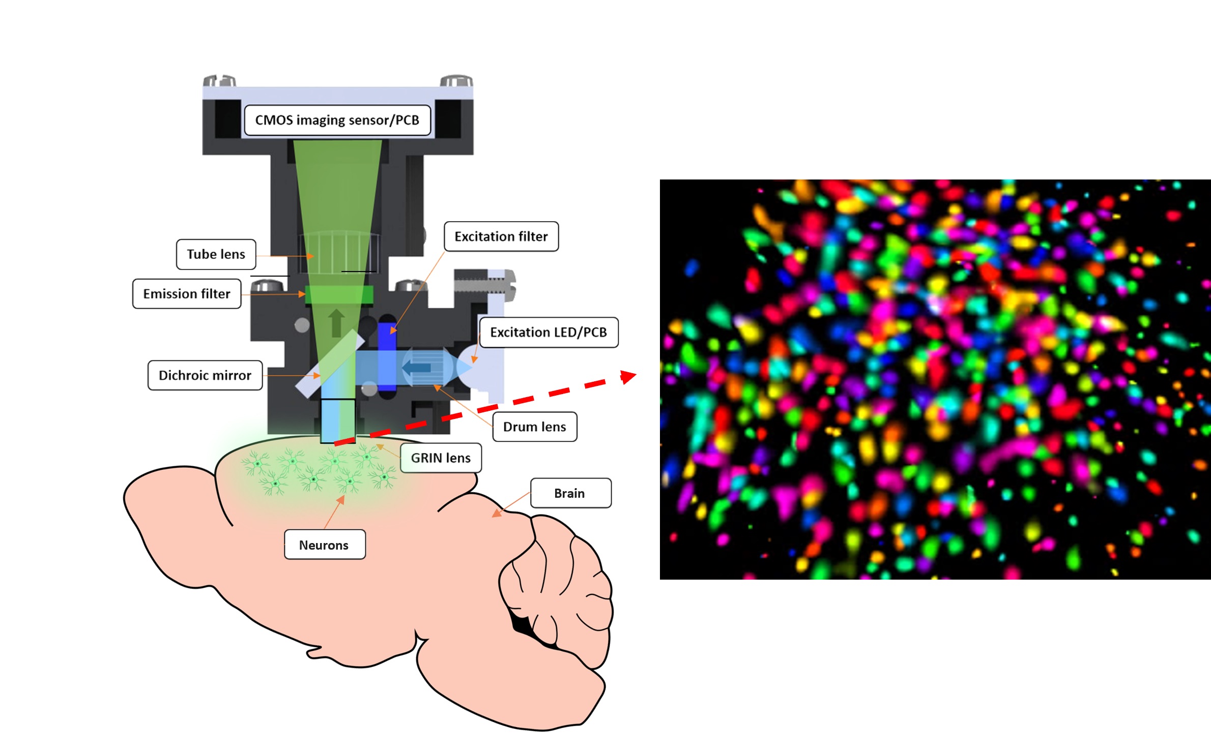
It involved electronics graduate research student, Dechuan Sun, implanting a light weight and removable miniaturised fluorescent microscope – called a miniscope – on top of a mouse’s head. This enabled us to observe brain cells in action in the mouse hippocampus, a region that encodes new memories and is strongly affected by Alzheimer’s.

Fluorescent microscopes work by detecting signals from Green Fluorescent Protein (GFP) connected to a calcium sensor molecule (known as GCaMP) that has been added to cells to ‘light up’ different cellular operations.
The discovery and development of GFP won the Nobel Prize for chemistry in 2008 and involves inserting a gene derived from a bioluminescent jellyfish into a cell.
To generate observable light signals when neurons activate, GCaMP DNA is inserted into the cells using a benign viral carrier, Adeno-Associated Virus (AAV). The neurons then generate their own GCaMP which changes fluorescence when neurons discharge.

The fluorescence changes because when a neuron activates, calcium flux binds to the protein and alters the intensity of the fluorescence. This activity can then be detected by shining light with the correct spectral properties onto the neurons.
This light comes from the miniscope.
The technology and technique for using small head-mounted miniscopes to monitor the brain was pioneered by Professor Mark Schnitzer at Stanford University.
Our miniscope weighs about three grams and has a small plastic lens about 1.8 millimetres in diameter that sits inside a tiny hole in the skull. The lens lies directly above the brain tissue – which means the brain tissue isn’t interfered with.
The images are then recorded directly onto a camera sensor chip similar to the ones in our mobile phones, and the data is then transported to a computer using a single one millimetre-thick coaxial wire.

The thinness and lightness of the wire is important to minimise any hindrance to the animal, but using such a thin wire requires some clever computer coding to compress the data.
The great advantage of the fluorescent miniscope is that it allows researchers to monitor activity in wide areas of the brain, enabling us to see how hundreds of neurons interact in an extended network – there’s no other practical way to do this.
And the fluorescent compounds can last for months allowing us to monitor animal brain behaviour over extended periods.

Health & Medicine
Meeting the challenge of dementia prevention in Australia
In our experiments, the mice were still able to ‘remember’ how to find the food reward on the linear track, but the miniscope showed us that the memory signals the mice were receiving were impaired.
More research is needed to more fully understand what’s going but this work could possibly illuminate a pathway towards finding restorative memory drug therapies.
It may also point the way towards being able to ’resynchronise’ brain signalling to reduce the noise caused by Alzheimer’s, possibly through techniques like deep brain stimulation.
According to the World Health Organization, more than 55 million people are living with dementia worldwide, with Alzheimer’s representing up to 70 per cent of those cases. And this number is rising as the population ages.
By 2030, the number of cases is expected to reach 78 million and by 2050 – there will be 139 million.
And this tells us that the need to better understand Alzheimer’s will only become more urgent.
This research was funded by the Australia Research Council under Discovery Project (DP170100363) and the Royal Melbourne Hospital Neuroscience Foundation.
Banner: Getty Images