Organoids: the next revolution in human biology has begun
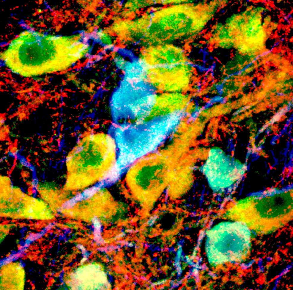
Researchers are growing miniature immature organs in dishes, meaning they can investigate a range of developmental disorders like epilepsy, autism and Alzheimer’s outside the human body
Published 14 October 2015
University of Melbourne biomedical scientists have joined the latest revolution in treating human diseases by growing tiny, immature organs, known as organoids.
These lentil-sized balls of cells, which scientists can grow in a dish, resemble the features of our brains, livers, guts, kidneys, prostates and pancreas, with the list rapidly expanding.
Organoids are creating huge interest because it means we can now study our biology in action outside a patient’s body. They are already being used to understand developmental disorders like epilepsy, autism, Alzheimer’s, Parkinson’s and a range of cancers.
Currently the organoids can’t differentiate into mature organs. They reach a certain size and start to die from the inside because they don’t yet have a fully functional blood vessel system for a continuous supply of nutrients and oxygen. But they already have enormous potential both as a research and therapeutic tool.
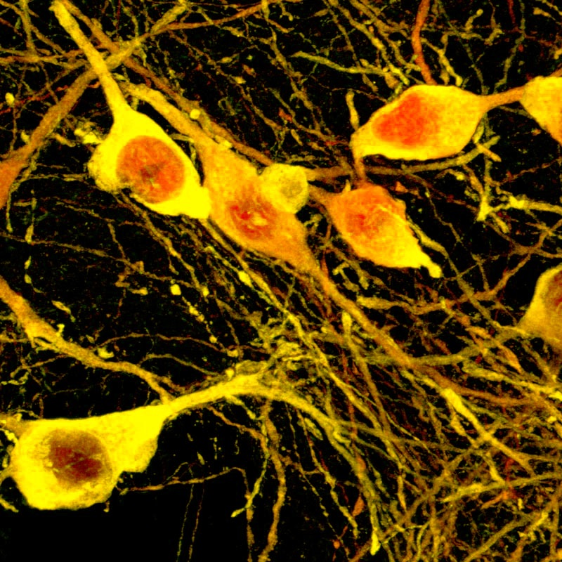
University of Melbourne’s Professor Melissa Little from the Faculty of Medicine, Dentistry and Health Sciences heads the Kidney Research Laboratory at the Murdoch Childrens Research Institute, The Royal Children’s Hospital. She is a world leader in kidney organoids and is working on childhood kidney cancers.
“Our kidney organoids are about 5-6 millimetres across, each has about 100 filtering units, and is starting to form blood vessels,” says Professor Little. “It’s still a very long way before a fully-fledged kidney but it’s a step in the right direction.
They may one day lead to rejection-free transplants for patients, once you have edited any inherited kidney disease genes.
At the moment the research team is generating little kidneys to use as a model to understand the mysteries of the disease mechanism.
“We are on the cusp of the revolution of understanding and treating disease,” says Professor Martin Pera, head of a large consortium of researchers at Stem Cells Australia, based at the University of Melbourne.
“Stem cell products are being used in trials for macular degeneration and spinal cord injury and some are being trialled as organ grafts for spinal cord injury and other diseases,” explains Professor Pera.
“The hope is we’ll eventually be able to scale up stem cell technologies to engineer or 3D print an entire organ.
“One of the most exciting use of organoids is to streamline the process of clinical drug trials. Currently drug trials are very long, extremely expensive, poorly predictive, and risky. Scientists are already using organoids to test drugs for efficacy or toxicity.
No more guinea pigs, we have organoids!
So how do you grow an organoid? First, you harvest skins cells from an adult human, reprogram them with protein signals to reverse the cell back into stem cells, the self-renewable “vanilla” cell type.
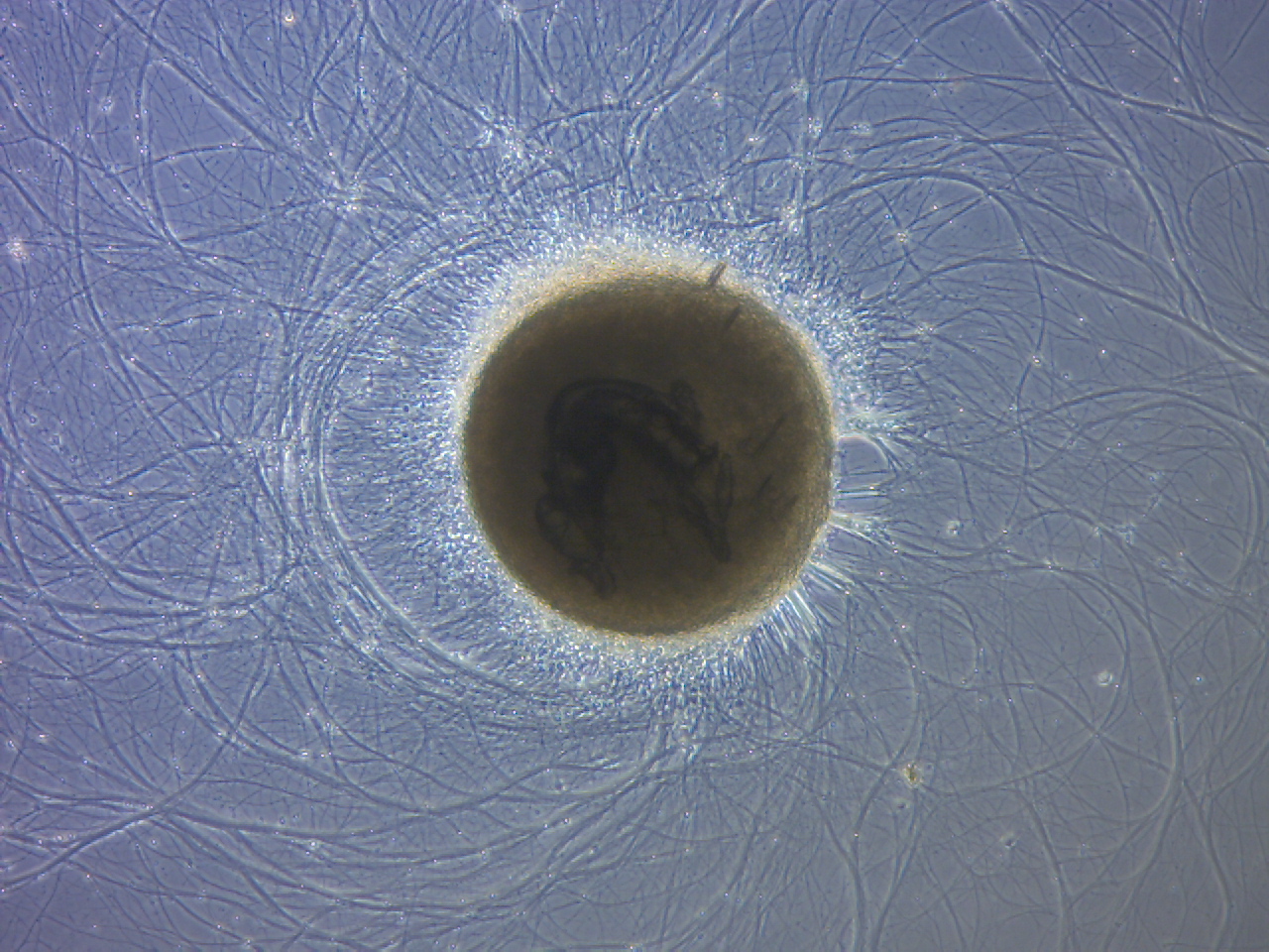
Then you biochemically prod the stem cells with specific growth factors to divide and develop them into a certain cell types, like brain nerve cells. Given the right growth factors at the right time and a nutrient oxygen rich environment, they will divide and grow into a brain organoid.
Dr Madeline Lancaster, the Austrian scientist who first grew organoids, noticed the cells in her tissue culture didn’t remain flat on the dish: instead they started to bob up, clump and reorganise themselves into 3D balls.
She decided to encourage the cells to grow. Analysis under a microscope revealed something remarkable, there were different types of brain nerve cells including immature retinal cells and early hypothalamus memory cells. These cells connected and self-organised to develop tissues almost like they do in a first trimester developing embryo.
So how do we study something as complex as autism or epilepsy using organoids?
The University of Melbourne’s Dr Mirella Dottori from the Centre for Neural Engineering, who is growing and studying organoids to understand autism, explains.
“The question for neurological disorders like autism is: where and when does the autism start? Does it begin in utero? Is it driven at all by genetic controls during brain development or is it something environmental during pregnancy or even later?
“Organoids enable us to study the types and proportions of various neurons and how they grow, migrate and connect with each other in 3D. Using this system, we can then make comparisons with organoids derived from autism and non-autism (‘control’) stem cells.”
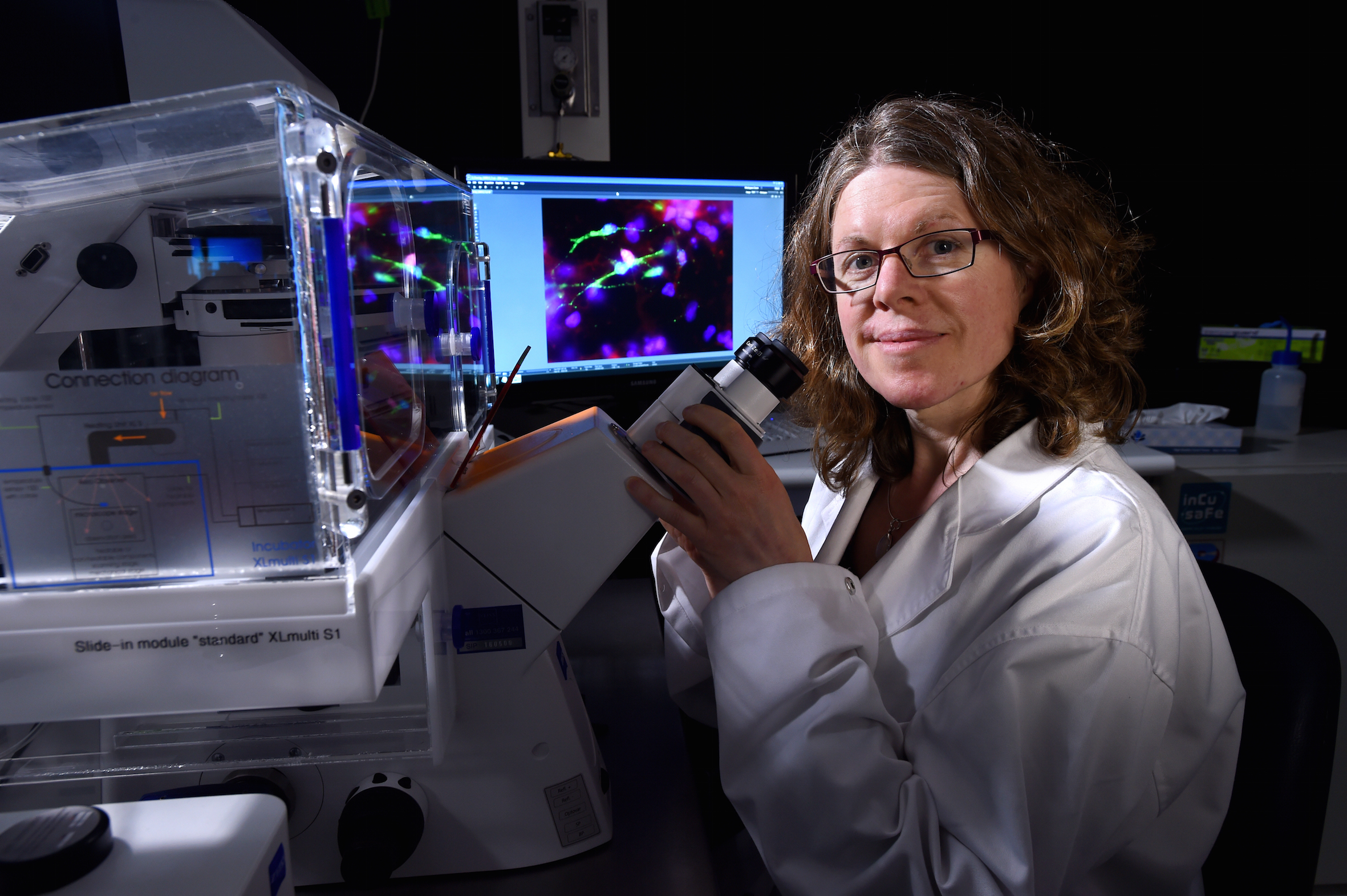
This means researchers can model questions about brain structure and function in the dish, by introducing various proteins, analysing specific nerve types and connections, and using electrodes to see how they are firing: something that cannot be easily done in human patients.
“Organoids are different to a single layer cell culture because in organoids the cells form 3D structures to ultimately form network connections,” says Dr Dottori. “It’s very exciting. They can grow to mimic specific parts of the brain. It’s so nice when we see something that we know goes on in the brain happen in the dish.”
This is the long awaited fresh approach to autism we need.
Dr Mirella Dottori is working with Professor Stan Skafidas and Dr Giovanna D’Abaco from the University’s Department of Electrical and Electronic Engineering, to develop a special 3D scaffold to not only extend the life of these organoids, but to also use inbuilt electrodes to measure nerve connections and activity.
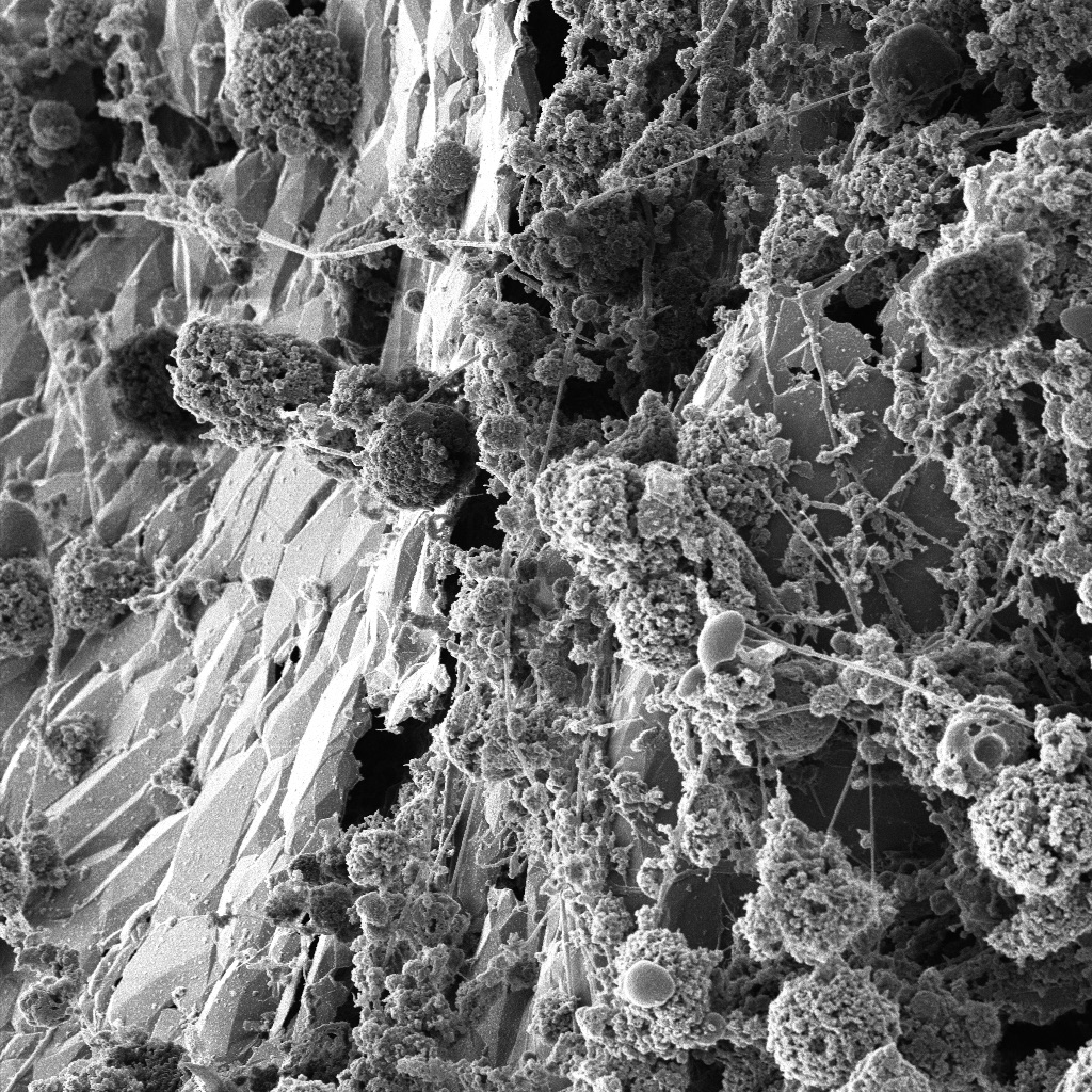
“Remember the organoids are very immature cells. If our team can extend their lives on, say, a scaffold, we have the opportunity to look at more mature cells. That would be really valuable.”
Professor Steve Petrou, Florey Institute of Neuroscience and Mental Health, says organoids enable scientists to explore new ways to treat neurological disorders such as epilepsy by modelling the condition in a dish.
“Epilepsy is a difficult disorder to study in animals and models, and experimenting on humans is unsafe and difficult,’’ Professor Petrou says.
“There is a severe form of epilepsy called Dravet Syndrome that afflicts children. The epilepsy manifests in developmental delays, intellectual and motor disability. Tragically they also can die young. In the last five years science has identified the genetic mutation and the markers, so we can now target our therapies.
“‘In the past we were treating the seizures with drugs, treating the symptoms and not the underlying cause. But the further you go back in the disease’s cellular pathway, the more chance you have of capturing the whole disorder.’’
Professor Petrou’s research is novel, essentially using the vast array of known drugs used for other medical conditions to see if any drugs can help those with this particular epilepsy.
The side effect of many drugs sometimes have unintended consequences when they act on other parts of a body’s system. Scientists aim to use this information to repurpose these drugs.
We plan to use organoids, that more accurately reflect the human diseased brain, to repurpose drugs expanding upon our current efforts using simpler model systems.
“Although we have had some success using simple models to show that the heart drug Quinidine can restore one aspect of the normal action of the mutated gene in epilepsy, there is urgent need for more complex genetic models to understand other aspects of the disease process.
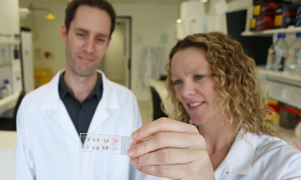
“Used in this way organoids should increase the rate of discovery of effective drugs to bring near term benefit for patients with previously untreatable severe epilepsy.”
If the revolution of human genome project provided a road map to study human diseases, then one could say the revolution of organoids is allowing us to analyse and eventually manage the traffic on these roads.
Banner image: Neurons derived from human stem cells connect to and talk with one another in the dish. Picture: the Florey Institute of Neuroscience and Mental Health


