Health & Medicine
Gene genies: Meet the researchers mapping our DNA to combat cancer

Researchers have unpicked our decades-long understanding of how babies develop in utero, revealing more about how birth defects may occur
Published 18 May 2018
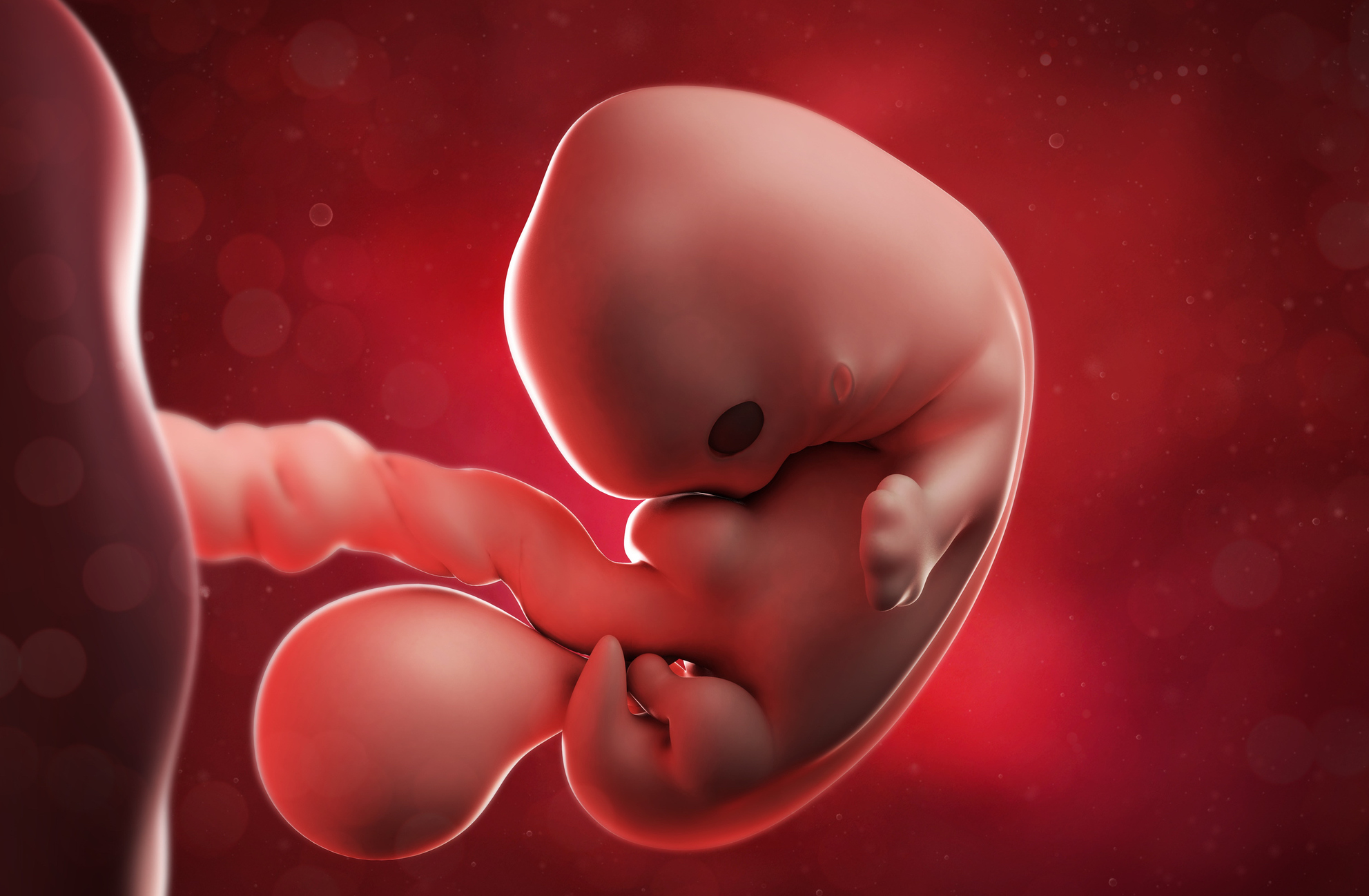
Since the 1940s, scientists have thought that programmed cell death, in which the body rids itself of unneeded cells, plays the main role in how babies develop in utero. Known as ‘apoptosis’, cell death is a normal process; driving, for example, the loss of our embryonic ‘tail’ at around 8 - 9 weeks.
But a new finding sheds light on how birth defects may occur. Researchers at the Walter and Eliza Hall Institute (WEHI) and the University of Melbourne have discovered that contrary to expectation, apoptosis plays a less extensive role than previously thought, and in very rare cases, embryos could still survive without cell death, although with severe defects.

“This challenges many of our basic assumptions about embryo development, and means we now have the potential to learn a lot more about many common birth defects, like cleft palate and spina bifida,” says Dr Francine Ke, a postdoctoral fellow at WEHI.
Dr Ke and her colleagues were able to pinpoint which tissues and organs depend on apoptosis to develop normally, like the palate in the roof of the mouth, and our fingers.
Other processes that relied on apoptosis included proper closure of the neural tube (that part of an embryo where the brain and spinal cord develop), and the normal development of major heart vessels.

Health & Medicine
Gene genies: Meet the researchers mapping our DNA to combat cancer
The team’s discovery came from their research into isolating the genes responsible for apoptosis.
Previous studies had already identified the genes BAK and BAX as playing a key role in programmed cell death. But when other researchers removed these genes in mice, they found that a number survived to adulthood, but with abnormalities like webbed ‘fingers’ and ‘toes’.
“This led us to think there must be another gene that steps in to fill the gap in the absence of BAK and BAX, to keep cell death functioning” says Dr Ke.
They suspected the lesser-known protein BOK, which had received much less research attention. An early step was to visualise it, to see how BOK’s structure compared to BAK and BAX.

They used a technique known as X-ray crystallography, where scientists grow a crystal of a complex molecule like a protein, then use advanced X-ray techniques to determine its structure. It’s the method used to build the commonly seen ball and stick representations of chemical structures.
The team used the Australian Synchrotron in Melbourne, a giant particle accelerator, which produces high-powered X-rays, to build a detailed atomic structure of BOK.
“This allowed us to determine the exact structure of the BOK protein,” says Dr Ke. “We discovered it is very similar to BAK and BAX, making it a good candidate for being the missing gene responsible for apoptosis.”

Health & Medicine
An unexpected step in the fight against stomach cancer
To test their hypothesis, the team removed all three genes – BAK, BAX and BOK – in mice to examine the impact on normal development.
They found that apoptosis was completely absent in these models, and this resulted in multiple abnormalities like spina bifida, exencephaly, heart vessel defects and cleft palate.
However, the team were surprised to find a majority of organs and tissues developed normally without programmed cell death.
“Though the embryos did exhibit abnormalities, they were clearly limited to particular organs and tissues. This implies that although apoptosis is crucial for normal development, the requirement for apoptosis was not as widespread as we had imagined,” says Dr Ke.
“Over the past 70 years, many studies have suggested that apoptosis was essential during the early stages of development, and in ‘hollowing out’ of body cavities and ducts in internal organs.

Health & Medicine
The simple, ethical case for gene editing
“However, we have now defined the exact tissues that genuinely require apoptosis.”
This new understanding will shape research into the formation and development of an embryos and the occurence of birth defects.
“It’s an important springboard for further research into devastating birth defects like spina bifida,” says Dr Ke.
“We’ve reframed some of the assumptions underpinning our understanding of these congenital birth defects, and that’s an important step towards finding ways that may prevent them in the future.”
The team, including colleagues Dr Hannah Vanyai, Associate Professor Anne Voss, Professor Andreas Strasser and Dr Angus Cowan, published the results in the journal Cell.
Banner image: Shutterstock