Sciences & Technology
3D scanning reveals new (but extinct) star fish
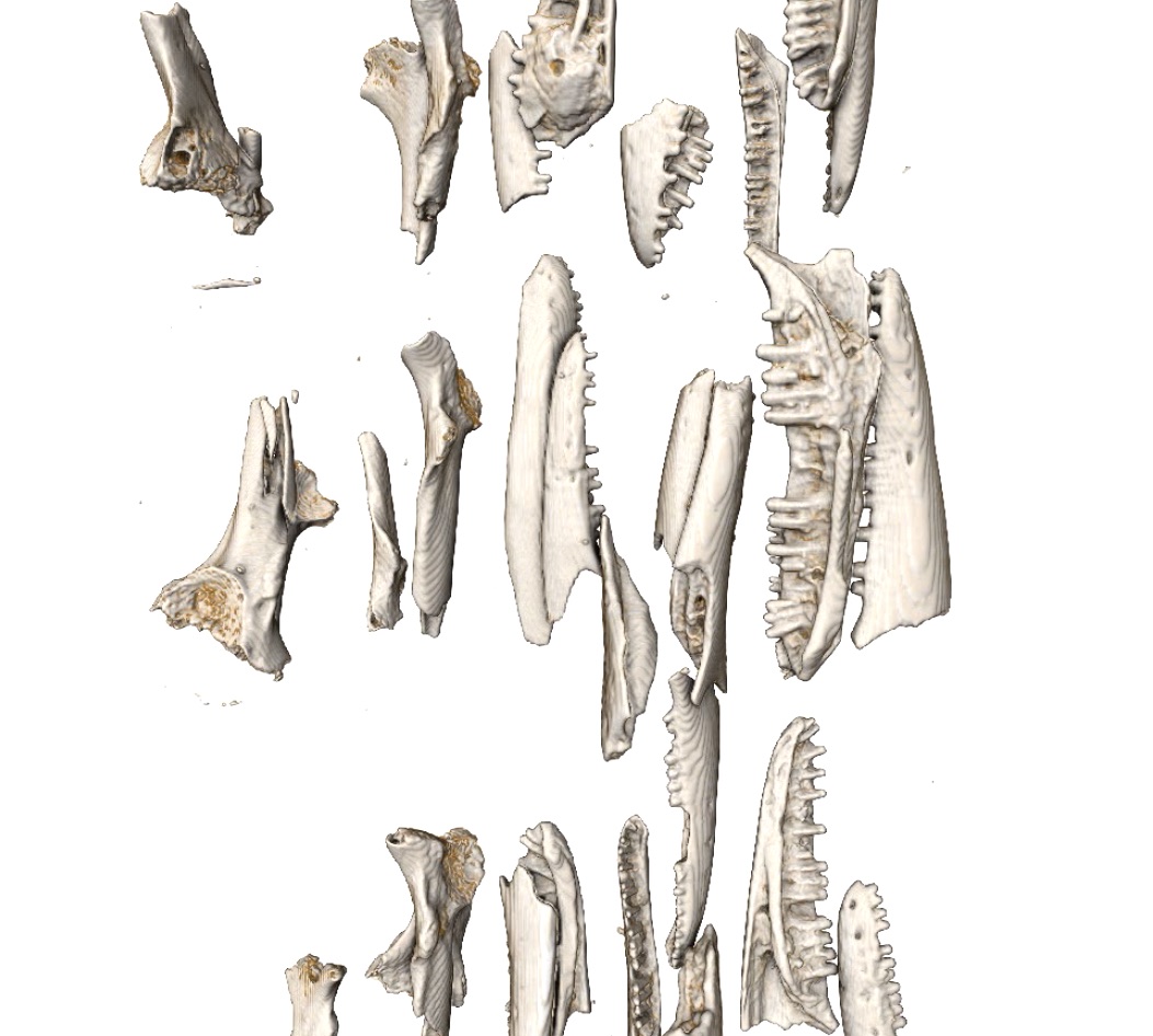
Highly detailed CT scans of the tiny fossilised bones of Australia’s lizards and frogs are telling scientists an important story of adaptation and extinction in a changing climate
Published 20 August 2020
It is no surprise that Australia is sometimes called “The Land of the Lizards”.
DNA analysis has revealed hundreds of new lizard species in the last decades, with more being constantly discovered. This extraordinary diversity, with nearly 900 species and counting, suggests a complex evolutionary past.
Yet we have little understanding of when, how or why this great diversity came to be.
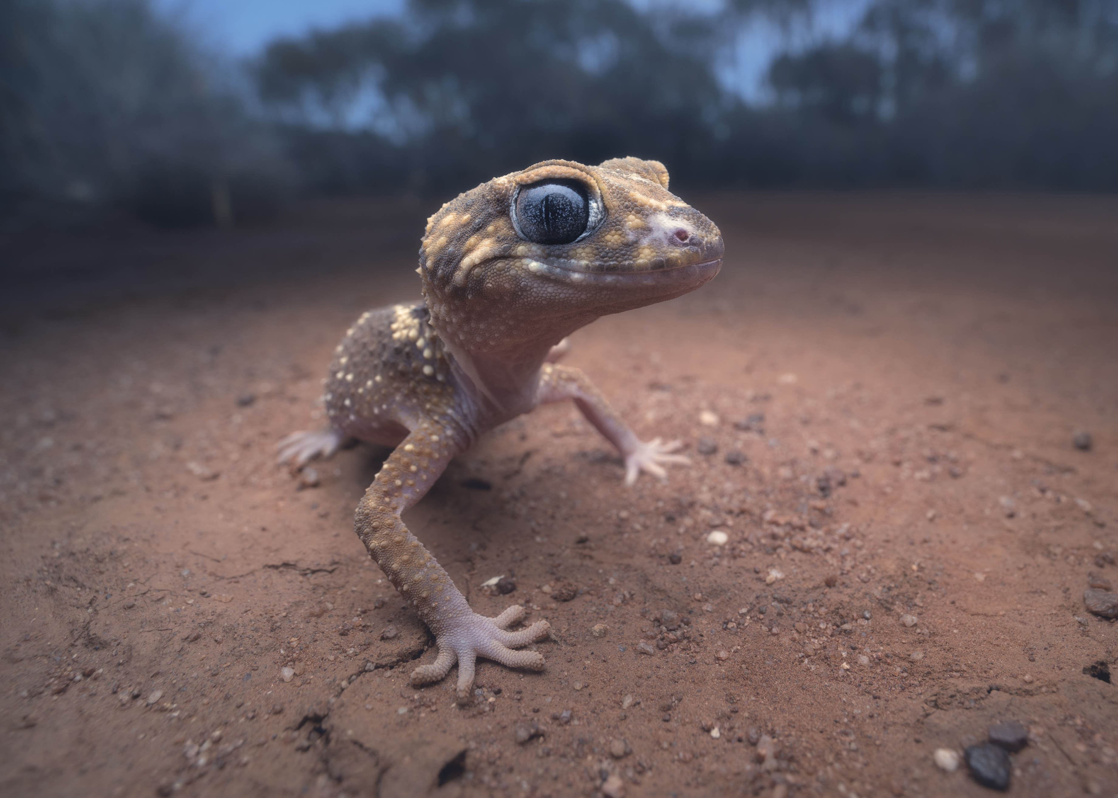
A lack of a fossil record is mostly to blame. With the exception of goannas, that once grew up to six metres long, these mostly small animals leave only tiny fragments of their bodies behind when they die, making fossilisation an unlikely prospect.
But, in some circumstances, when particular conditions are right, these tiny remains fossilise and can be preserved for millions of years underground.
A major challenge for palaeontologists, whose job it is to identify and interpret fossils, has been to use these tiny remains to learn more about Australia’s natural history.
This is important because understanding the contexts that generated evolutionary diversity in the past may help to reveal how species will respond to climate change in the future.

Sciences & Technology
3D scanning reveals new (but extinct) star fish
Many of Australia’s reptiles and amphibians, known collectively as herpetofauna, are threatened with extinction due to global warming, habitat loss and invasive predators.
They have adapted over millions of years through past climatic changes, including intensifying aridity, Ice Ages and the arrival of Asian species.
Much of the herpetofauna found here in Australia aren’t found anywhere else.
Our knowledge of Australia’s recent prehistory is largely focused on our cute and cuddly creatures – the mammals – including marsupials and monotremes. But, it’s our lizards and frogs that boast greater species diversity and can tell us more about how animals adapted to or succumbed to past environmental upheaval.
To identify the conditions under which these animals evolved, we need to compare the past herpetofauna with those from the present day.
Fossils offer a unique opportunity to do that, providing direct evidence of what kinds of species existed before major environmental change compared to those that survived it.
But to do so, we first need to know what species the fossils belong to. And for this we need teeth and bones – lots and lots of them.
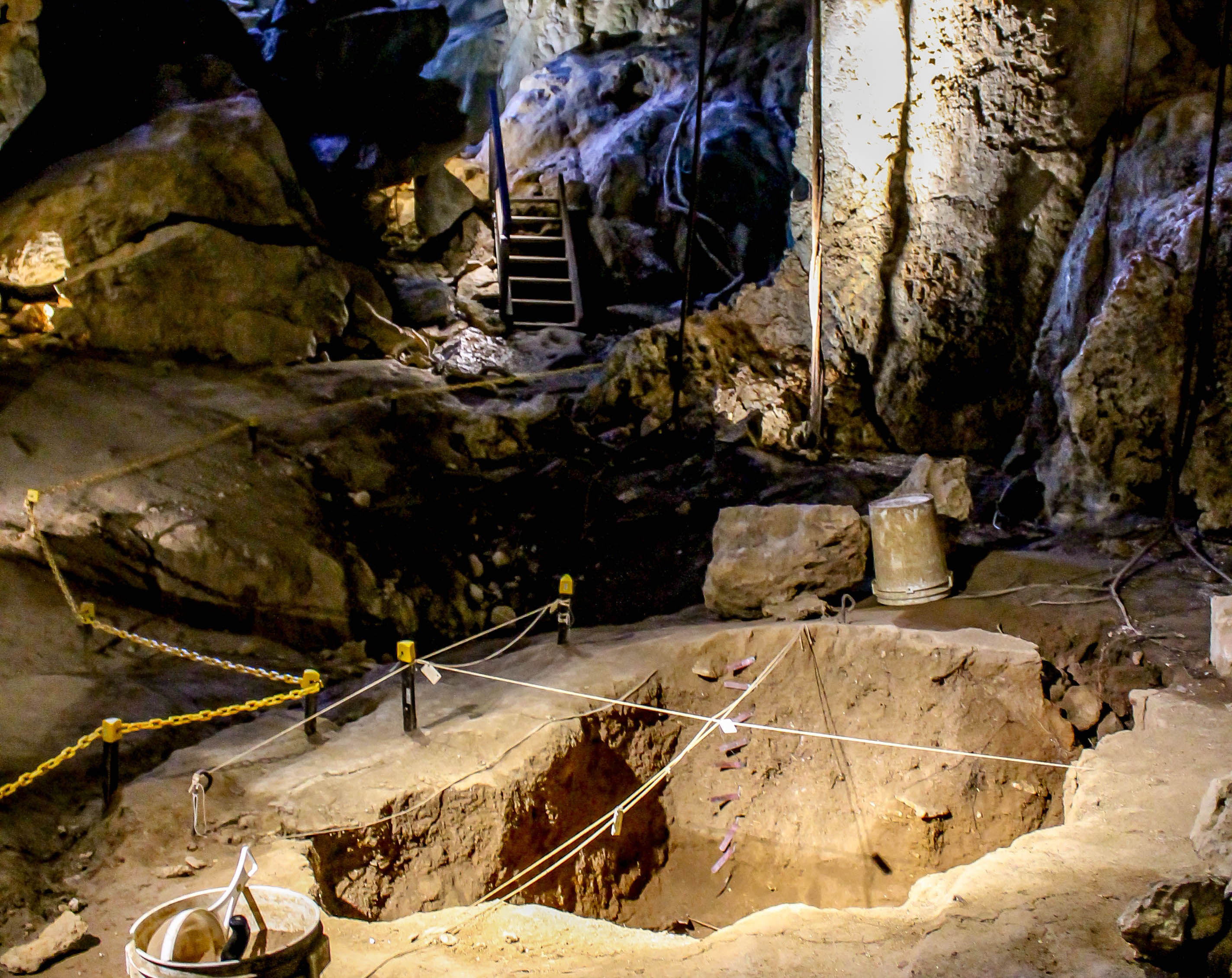
Thankfully, owls and ghost bats that roost in caves have been doing us a favour by capturing lizards and frogs as their prized food, night after night, for millennia. The bits they don’t end up eating fall to the cave floor and, over tens of thousands of years, these layers of bones accumulate into a nightly record of life, preserved for palaeontologists to unravel.

Each bone tells a story of who that individual was and who it lived alongside – each bone is a data point into the past. These fossils, now carefully housed in museum collections, are the fruits of countless hours of excavating in places like Capricorn Caves in central-eastern Queensland.
However, piecing together an accurate story from these fossils is difficult, because not only are they small and broken, we also have thousands of them to sort through and identify.
OLD MATERIAL, NEW TECHNOLOGIES
This is where our newly developed high-throughput method comes in.
Using simple, inexpensive materials like pharmaceutical capsules and paper straws, we can secure hundreds of tiny fossils in a single tube so we can scan them together using X-ray computed tomography (CT scanning).
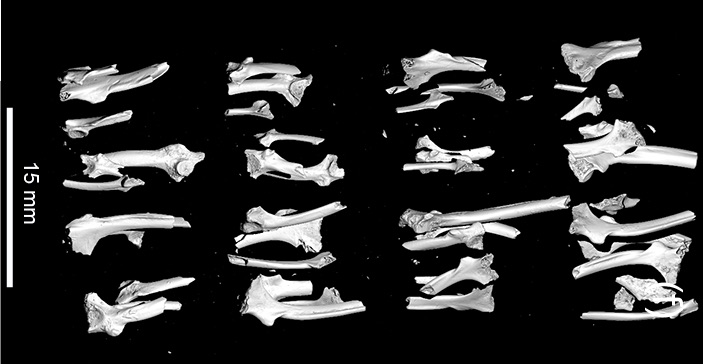
This involves taking thousands of X-rays of an object and combining them into a 3D volume. Although CT technology, called microCT for very small objects, has existed for decades, it’s been troublesome to scan many samples at once.
Each specimen must remain still while being X-rayed and then be individually identified and labelled amongst dozens of other fossils.
Using our new set up, we can now scan entire collections of tiny fossils, creating 3D images with a resolution of around 20 micrometers or less. That means each pixel (or voxel in 3D) shows details that are less than the width of a human hair.
You can see and interact with these 3D models and more here.

Sciences & Technology
Australia’s mountains are still growing
These scans also reveal internal structures like the roots of teeth, which would otherwise be hidden inside the jaw bone. That’s a major advantage for fossils, since palaeontologists generally don’t want to destroy them by cutting them up.
We will now use these 3D images to compare the fossils with their modern relatives – also CT scanned from museum collections – to identify the impacts of climate change on Australian herpetofauna.
In this way, we can study whole communities of lizards and frogs to see how they have changed over time – whether they stayed in place, adapted to new conditions, moved elsewhere or gone extinct.
To draw these conclusions, we need to compare hundreds of specimens, checking sizes, shapes and even the differences between males and females before we can be certain who they are. We can’t do this by eye, so we use sophisticated software to quantify complex morphologies.
Generating 3D models of fossils allows us to study them virtually without the need to touch them – which would otherwise risk potentially breaking and losing them forever.
This is the ultimate insurance policy against loss, because if we have a digital replica that is highly accurate – so we have preserved the specimen in more ways than one.
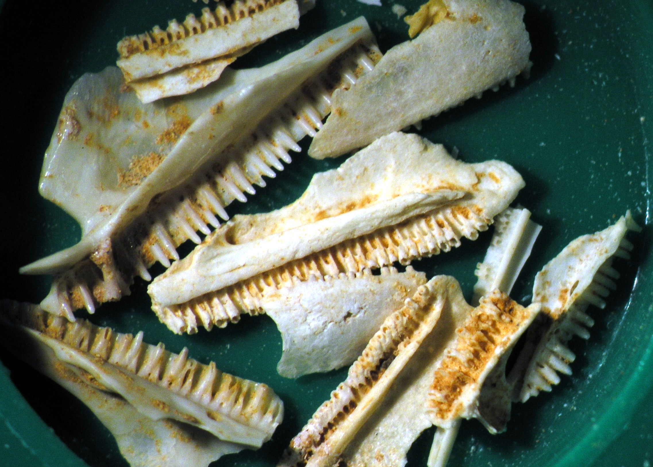
We can also apply new techniques to these bones to determine not only the species of lizard or frog but also how it lived – how it moved around its habitat, how big it was, did it suffer any diseases and, most importantly, how many species lived in one place at one time.
For example, finite element analysis (FEA) uses principles of engineering to model stresses and strains across a structure related to biomechanics, which in turn can tell us if the animal likely swam, hopped or climbed, or even what type of food it ate.

Sciences & Technology
Going beyond political borders to protect threatened animals
Armed with these digital models we can collaborate globally. This means that scientists working on similar questions across the world can see and interact with precious specimens, without the risk of them being lost or damaged in transit.
Finally, we can display these specimens to educate the public on their importance in ways we couldn’t do using the fossils themselves. They are simply too small and delicate to handle.
By 3D printing enlarged versions of them we, as scientists and science communicators, can teach students about the history of life on earth, Australia’s natural heritage, and what we can do to protect it.
The tiny bones of these creatures have been scattered and buried over millennia. It’s time we allowed them to tell their stories while resting in peace.
Rocio Aguilar, research fellow at Monash University and research assistant at Melbourne Museum, is a co-author of this research.
The authors thank the Melbourne TrACEES platform for access to the micro-CT scanner used in this study.
Banner: CT scans of frog and lizard fossils/ Supplied