Arts & Culture
Bones of contention

A collection of leg bones at the Melbourne Dental School was originally intended to play a key role in forensic medicine, but unexpected discoveries along the way have opened up whole new areas of research from bone strength to ageing
Published 30 June 2017
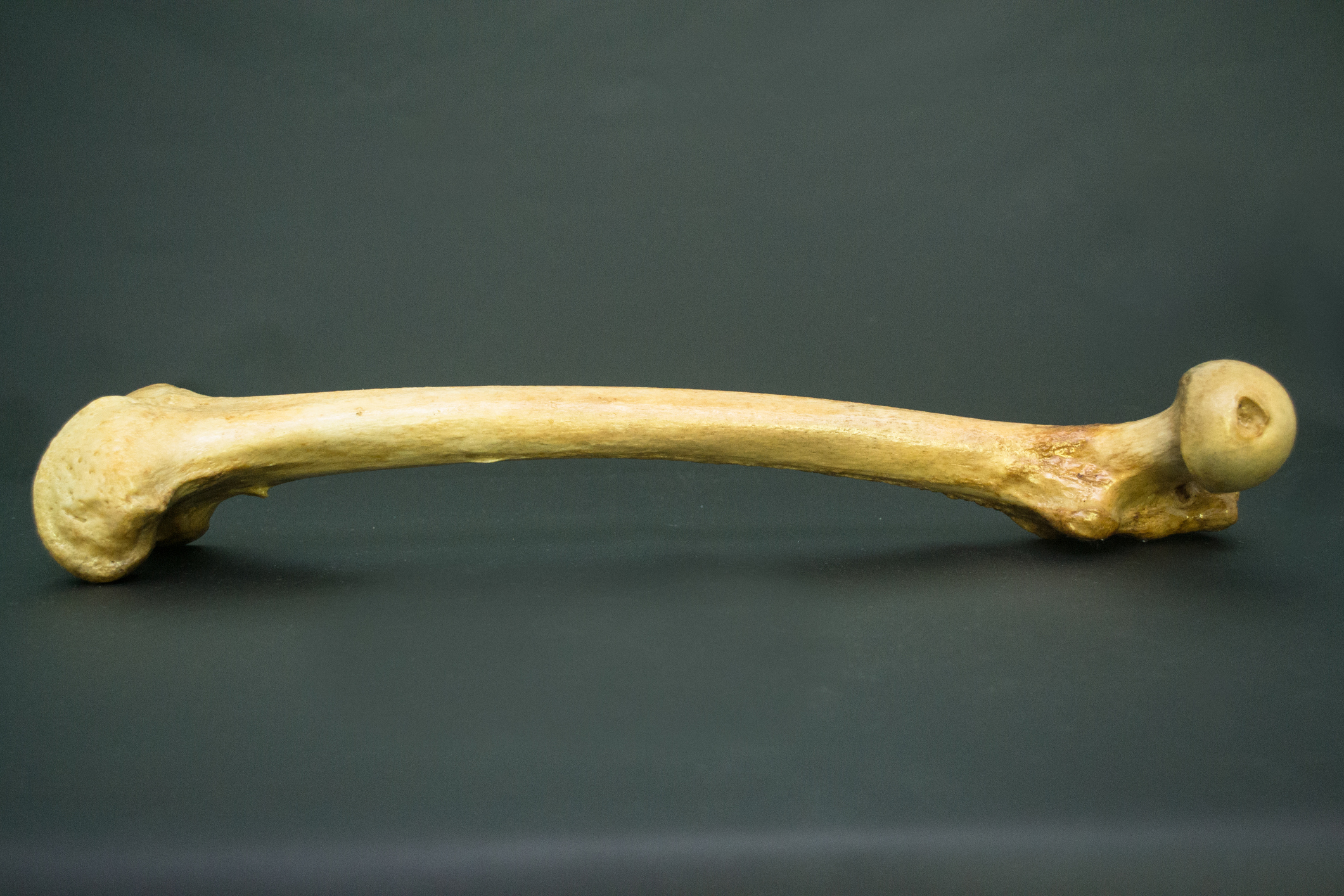
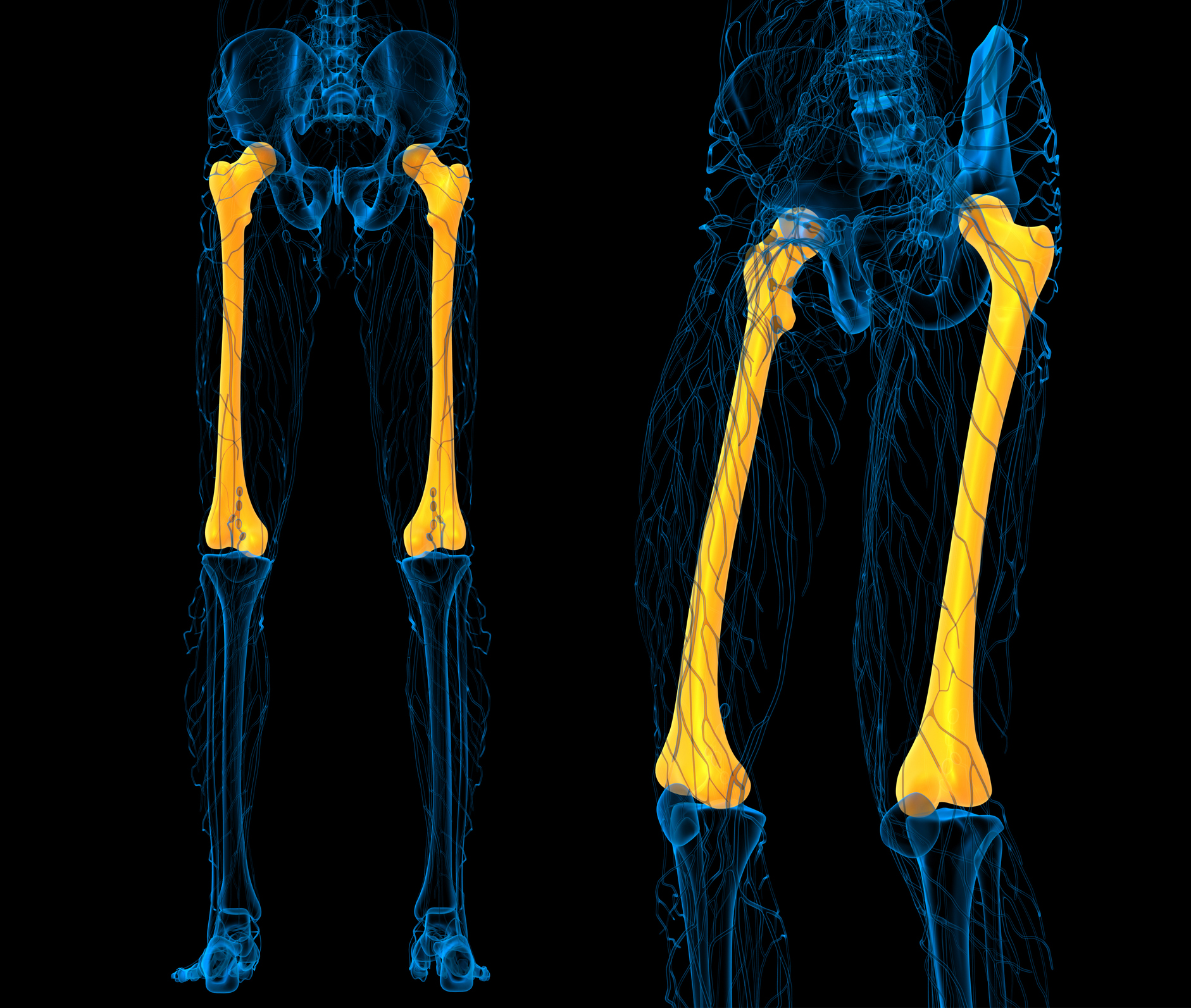
The most durable bone in the body is the mid-shaft of the femur, or thighbone.
In nature, when bodies are left unburied, the elements eventually expose the bones and they begin to break down. Scavenging animals may speed up the process, rapidly destroying much of the skeleton. But in both cases, it’s the middle of the thighbone – which is very thick - that most often survives and can be analysed.

While the connection between leg bones and dentistry is not immediately obvious, the femur’s attributes are the reason the Melbourne Dental School holds over 600 specimens in its Melbourne Femur Collection: a collection attracting the attention of researchers from medical science to anthropology internationally, but with origins in forensic dentistry, or forensic odontology.
Forensic dentistry deals with teeth, and the marks left by teeth, to infer the history of a person. It is an expertise many of us have seen dramatised on TV when dental records prove central to the identification of a victim, or one bite mark solves a murder. But there are other critical applications of forensic odontology, such as in the aftermath of natural disasters or war and rapid and effective identification is critical for both families of victims and legal redress. It is also critical in anthropology and archaeology in helping reconstruct our deep past.

Arts & Culture
Bones of contention
In 1989, forensic odontologist Professor John Clement, founder of the Melbourne Femur Collection, arrived at the University of Melbourne.
When faced with one of the central problems in forensics – establishing the age at death of human remains – Professor Clement, now the Melbourne Dental School’s Director of Research and Deputy Head, went straight to the femur.
At the time, the sequential emergence of teeth until adulthood could be reliably used to establish the age-at-death of younger human remains. But once all the adult teeth have come in there wasn’t a way of confirming age-at-death through teeth alone.
The femur, with its thick cortex – the outer layer of the bone – was the perfect candidate. As we age our bones start to thin and there are changes in their microstructures. It seemed that science might be able to tell the age at time of death by examining the cortex. Presumably, in younger bones the cortex would be thicker and denser and in older ones, thinner and more porous.
The story however proved to be much more complicated.
During this early research to determine age-related changes to bone, the Melbourne Femur Collection emerged. Professor Clement and his team worked with the strong support of the Victorian Institute of Forensic Medicine (VIFM) and, with the generosity and trust of donor families, developed a collection whose provenance was impeccable.
They knew exactly where the bones had come from. They had been screened for diseases that might affect structure, and had come from a fairly narrow population. Factors such as poor nutrition were unlikely to skew results, and importantly, age at time of death was known.
In knowing age-at-death, researchers would be able to test whether there was a standard range in the structure and density of bone within each age group.
The results were emphatic. Across each age group there was an enormous variation in the thickness, density and microstructures of the cortex. There was no way the cortex could be used to reliably determine an individual’s age.
It was a disappointing revelation; and useless for forensics.

But the research was getting noticed in other circles.
In the mid-1990s, medical research into bones relied on x-rays that could only look at the gross structure, and mineral density could only be inferred through calculations. They couldn’t see the important finer detail inside the bone.
Meanwhile the team at the Melbourne Dental School was looking into the bone at increasingly fine levels of detail, using newly emergent technologies and approaches.
What became particularly apparent were the methodologies that dentists were bringing to the table – as specialists in hard tissue, or bone – and the care with which the collection was formed, resulting in trustworthy, robust data.
It got the attention of specialists in bone health and osteoporosis, who immediately saw the value and applicability of the collection and the research the dental school was doing.
So began the cross-disciplinary possibilities. Medical researchers, anthropologists and other scientists from Cambridge to Saskatchewan got in contact with the team, resulting in numerous significant international collaborations that have so far resulted in over 80 publications.

Health & Medicine
Legs, ligaments and longevity
In terms of bone health, the Melbourne Femur Collection opened up a whole field of inquiry, with implications for diseases that threaten to impoverish us all as the population continues to age, such as osteoporosis.
The strength of bone is not just reliant on its thickness, but also its density, and its porosity. A porous bone – essentially full of pores or holes – is weak. For a long time it was thought that hip fractures in older people were the result of falls. Recently however, it’s emerging that a slight knock against a table may fracture a weak hip, and the fall follows.
In your seventies, a hip fracture doubles your risk of dying within a year. For women 80 and older who are in excellent health, it triples.
With an increasingly ageing population, it promises to be a huge public health issue.
But, as the Melbourne Femur Collection helped establish, weaknesses in the bone are not uniform, nor inevitable, but individual. And the strength of an individual’s bone is dependent on critical periods, such as late adolescence, when we reach peak bone mass.
“If we know the optimal exercise and nutrition interventions at these critical ages,” says Professor Clement, “and target them, such as getting teenage girls to exercise on rowing machines – the perfect weight-bearing exercise for hip strength – we may be able to affect a generation of women, ensuring longer, higher quality lifespans.”
The impacts would be enormous.

Preventing a generation of osteoporosis may seem a long way from forensics. For the team at the Melbourne Dental School, it’s a possibility that couldn’t have been imagined when the collection began.
“It has a lot to do with serendipity,” says Melbourne Femur Collection Curator and Lecturer of Anatomy, Dr Rita Hardiman. “You don’t know what you might find.”
What the researchers first went searching for eluded them. Instead, by staying close to examining age-related changes to the bone, what started as a way to determine the age at death of individuals has in fact revealed findings of enormous relevance to us all.

With so many disciplines in the mix, the impacts may be felt from bone health to anthropology and archaeology, where findings may help us piece together the human story over vast timeframes.
“Blue Sky thinking?” Professor Clement asks. “If we grow the collection in the future, we’ll be able to assess and compare a whole lot of demographic information that reflects Australia’s changing population.
“With a lot more information we’ll be able to explore some very big questions, such as the effects of pollution on our bones and of climate change. We may be able to intervene in some of our most pressing bone diseases.
“But it’s only going to be possible through collaboration – by making the information and collection available to colleagues who can bring their expertise to the table.”
It’s collaboration that has always been at the centre of the collection and the research, from multidisciplinary connections to the relationship with the VIFM. And, at the heart of it all, the donors and donor families who have made it all possible.
“It’s humbling, and a great gift,” says Professor Clement. “It’s a custodianship which comes with ethical obligation to study the collection to the maximum benefit for those who are yet to come.”
Banner image: iStock